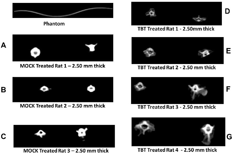
Fig. 2. Micro Computer Tomography Scanning (microCT scan) from vertebral bone of both TBT-treated and MOCK Treated rats. To make sure that both mineral and/or organic matrix were being affected by TBT we analyzed vertebrae from TBT, we evaluated the tri-dimensional cuts from vertebrae (one of the major sites of bone breaking in osteoporosis). A-C show four different TBT-treated MOCK treated at cut-offs of 2.5 mm thick transversal cuts were one can observed abundance of mineralized tissue (higher white signal - parameter used appropriate Phantom blocks of calcium carbonate rich seen below.). However, from D-G it is visualized less abundance of mineralized bone whereas sites of soft (less mineralized organic matrix) is still detected in TBT-treated animals (n=4). Quantification of these intensities in proper software indicated that with no doubt that TBT showed, at least in this part of the skeleton less mineralized tissue. Studies with other bones are still in progress.